Melanoma
Early-stage melanoma can be healed totally. New continuously developing medication brings a better prognosis even in advanced melanoma. Clinic Helena offers the best available treatment in melanoma care!
Melanoma starts in skin melanocytes (i.e. the cells that produce the dark pigment in the skin). Along with breast cancer, melanoma is the fastest growing cancer in the western world.
The main risk factor is the burning of the skin due to UV radiation from the sun. Pale and freckled skin with red pigment in it burns most easily. It is recommended that on sunny days, sun creams with a minimum sun protection factor (SPF) of 20 should be used.
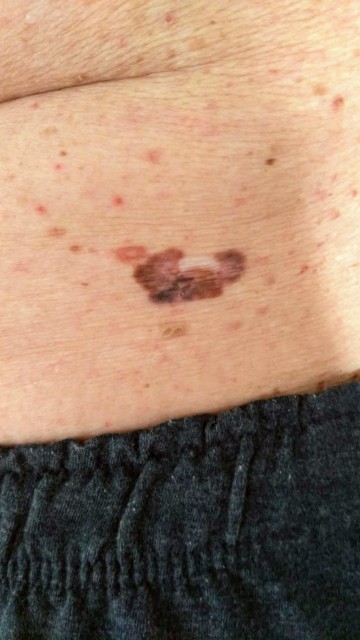
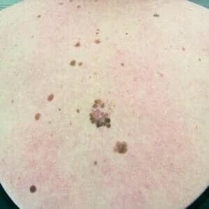
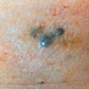
Melanoma develops in a mole of the skin in only about a third of the cases, whereas in two-thirds of the cases melanoma develops on previously intact skin. It is important to remove for sampling all moles and skin abnormalities if there is a change of colour or size, if there is redness around a mole, or if the skin surface is broken, develops a scab and will not heal.
A mole is removed completely for sampling and, if possible, a small margin of normal skin around it is also removed. All removed moles are examined under a microscope for pathoanatomical diagnosis (PAD).
Treatment
The primary treatment of melanoma is surgical excision. The extent of the excision is determined based on the thickness of the melanoma. The thickness is measured from the PAD biopsy and expressed in millimetres on the Breslow scale. Surface ulceration makes the estimation of the thickness more difficult, and as a rule the real thickness is greater than the estimated thickness. Melanoma spreads to its immediate surroundings forming so-called satellite metastases. The distance of these metastases from the primary melanoma depends on the thickness of the melanoma. Therefore, the amount of healthy tissue that is removed around the primary melanoma is determined on the basis of the Breslow thickness.
Breslow thickness of less than 1 mm indicates a superficial, early-stage disease with a good prognosis. In this case, 1 cm of healthy tissue is removed around the melanoma or the scar of an earlier excision.
Breslow thickness of 1-2 mm also indicates a superficial melanoma with a relatively good prognosis, and removal of 2 cm of healthy tissue around it is sufficient.
Melanomas with a thickness of more than 2 mm are removed with 2-3 cm margins of healthy tissue. Wider margins serve no purpose, and extensive tissue removal with 5 cm margins which used to be common is no longer used.
Sentinel node
The primary treatment of melanoma is surgical excision of the tumour. The surgical treatment of melanoma always includes a sentinel lymph node biopsy if the thickness of the melanoma is more than 1 mm.
On the day preceding the procedure, the melanoma scar is injected with technetium radioisotope, and lymph node mapping is performed. The sentinel lymph nodes that take up the radioisotope are removed in conjunction with the additional excision of the melanoma scar.
If in further examination melanoma cells are found in these detected sentinel nodes, lymph node dissection is performed to remove all lymph nodes in that area of the body.
The surgery can be performed as day surgery under local anaesthesia, but if the lymph node area is large the surgery will be performed under general anaesthesia. The patient can stay overnight in the postoperative unit if necessary.
At our clinic, the surgery is performed by a plastic surgeon using oncoplastic methods. Local skin flaps, free flaps and, if necessary, microvascular tissue flaps are used.
The cost of the surgery depends on the extent of the procedure and will be estimated at the consultation appointment.
Adjuvant therapies
If the thickness of the melanoma is more than 1 mm, our oncologist (Dr. Esa Männistö) will be consulted about the necessity of adjuvant therapies.
In order to find out the stage of the melanoma, an X-ray of the lungs and an ultrasound scan of the liver are taken. In addition, CT scans of the body, as well as a bone scan, are performed on patients with lymph-node positive melanoma.
In recent years, the drug treatment of melanoma has advanced significantly. As a result of studies of the role of mitogen-activated protein kinases (MAPKs) in the regulation of gene expression and of the growth and survival of cells, new tumour markers (BRAF Gene mutation analysis) have been identified. These are of assistance when new drugs are selected for the curative treatment of metastatic melanoma. The disadvantage of the new drugs is their high price.
Our oncologist Esa Männistö will recommend the best possible medication for each individual patient. Drugs that have been used for a long time, such as interferon-alpha (IFN-α), are still effective in the prevention of recurrence of the disease in certain cases.
Adjuvant therapies also include multi-cytostatic therapy, chemoimmunotherapy (combination of cytostatic and IFN-α therapy) and radiation therapy. Melanoma vaccines are still at the research stage.