Facial Reconstructions and Tumours
Eyelid reconstructions
Ectropion is a medical condition in which the lower eyelid loosens and turns outwards. In this case the lid does not protect the surface of the eye anymore. The eye is easily irritated and inflamed, and it becomes watery. The tear liquid tries to rinse the eye even more to repair the situation. An eye infection can damage the surface of the eye and lead to loss of eyesight.
In an ectropion surgery the eye lid will be shortened and fixed to the supporting tissue of the eye’s outer corner. Sometimes, the lower eyelid’s skin becomes scarred, and in these cases, healthy skin can be taken for example from the upper lid with a double side flap. In these cases, the surgery is performed in two stages, where first the skin loss is repaired and in 2-3 weeks’ time the inner corner flap edge is separated and finalised.
Entropion is a medical condition in which the eyelid turns inwards. The eye lashes irritate the surface of the eye. Again, the eye is easily inflamed and becomes teary. In the surgery excessive skin and tight scars are removed. Eye lid’s mucous membrane can be reconstructed with a mucosa graft from the mouth.
In case the whole eyelid needs to be removed due to a tumour, the skin is reconstructed with a local flap. The eyelid’s connective tissue (tarsus) is replaced with a cartilage graft, which can be taken either from the nasal septum or from the ear. A sleek mucus membrane from the mouth is often needed to avoid irritating the eye.
Nasal reconstructions
Rhinophyma is an unfortunate condition of overgrowth on the surface of the nose. The best form of treatment is to peel the surface away and to reconstruct it either with a skin transplant or forehead flap.
Cancerous changes and burn wounds may demand the whole nose to be reconstructed.
The whole external nose can be reconstructed for example with forehead flap. The nasal septum support can be taken from the pelvic bone (crista) and the cartilage from the ear. The interior wall skin of the nose is usually replaced with a skin graft. Functionally, the nose no longer moisturises the air breathed in, yet the aesthetical result is satisfying.
Ear reconstructions
In protruding ear, the outer ear development has been compromised and the so-called antihelix fold can even be totally absent. In the corrective surgery, the cartilage is folded into its normal shape. Operations on adults can be done in local anaesthesia. Children are usually operated on under general anaesthesia. After the operation, we recommend supporting the position of the ears by using a headband, for example, a normal sports band.
Microtia is a condition where the ear is partially or completely undeveloped. Often the patient is also lacking inner ear, so only the bone conducting hearing is available and can be used with specially constructed and implanted hearing aids.
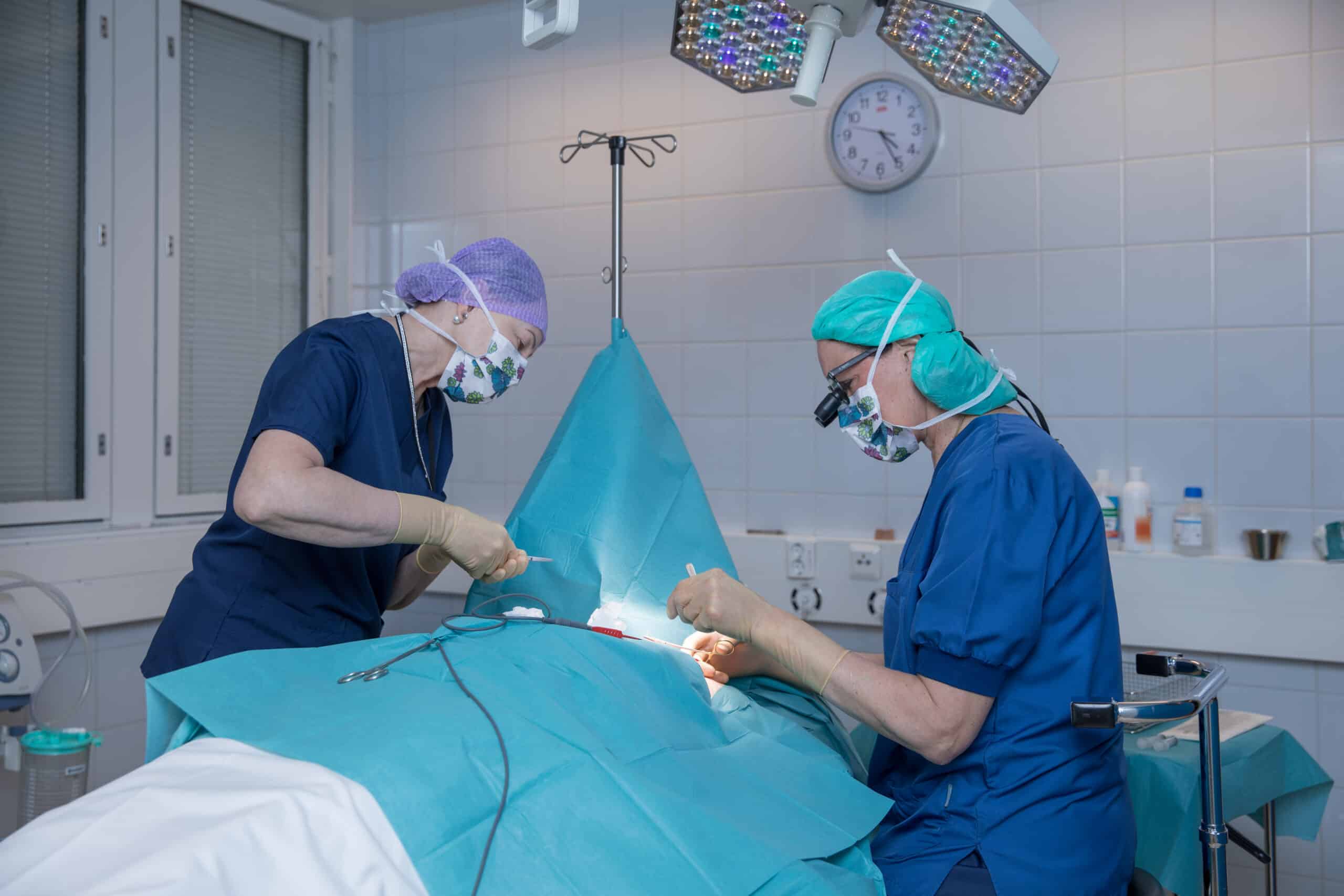
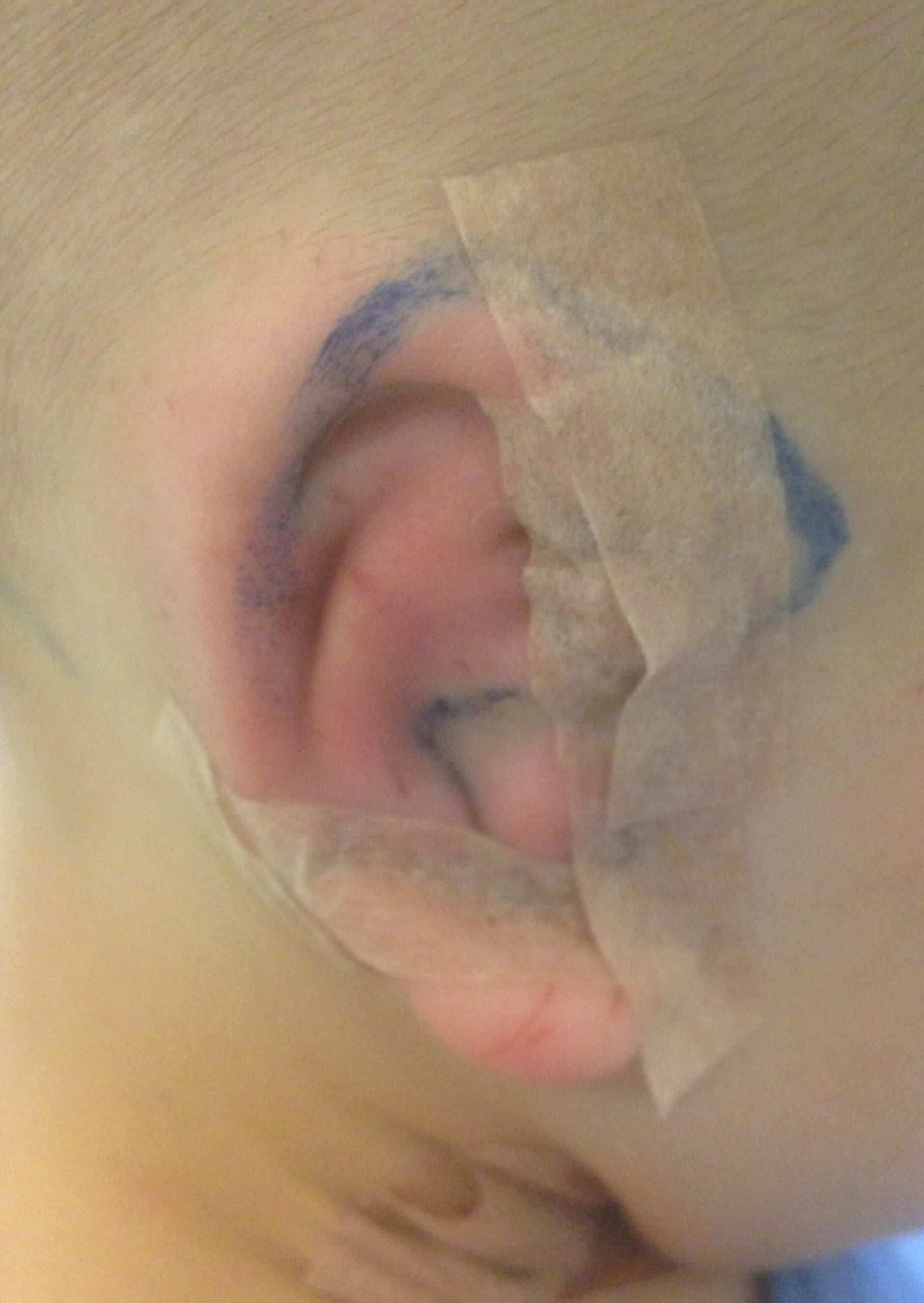
The outer ear can be constructed with own tissue rib cartilage or artificial material. An own tissue ear reconstruction is two phased. When the child’s head has grown almost into its full size by the age of 10, the ear mould formed of rib cartilage is placed under the skin in its own place. After the recovery, in about 6 months’ time, the ear is modelled.
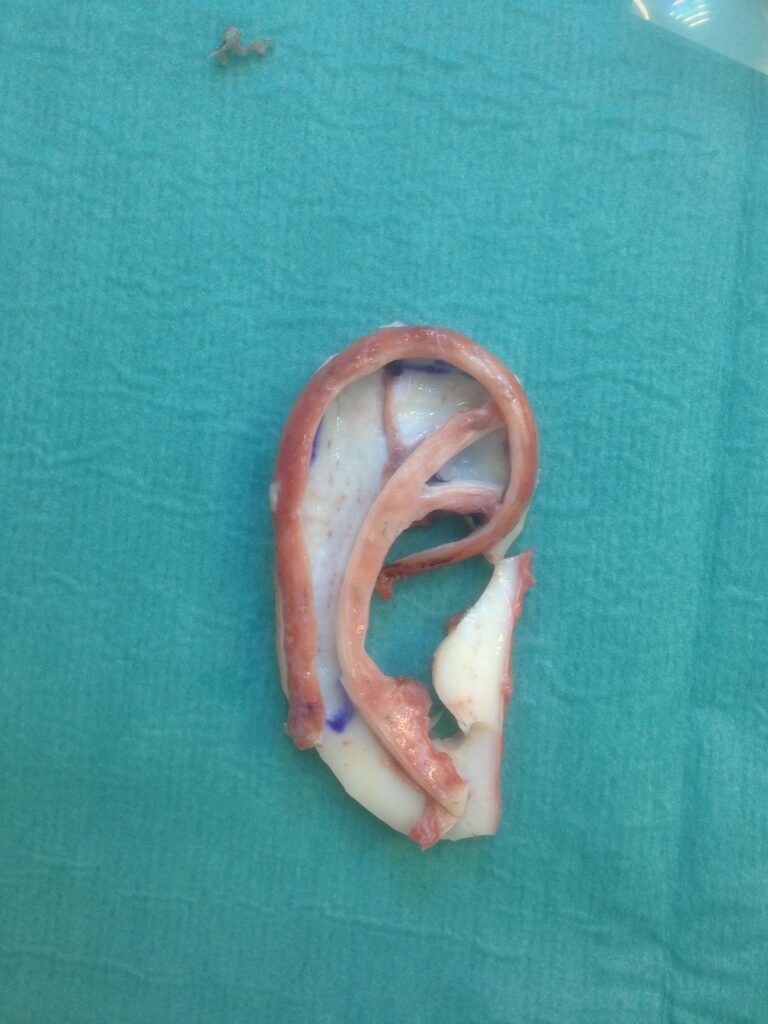
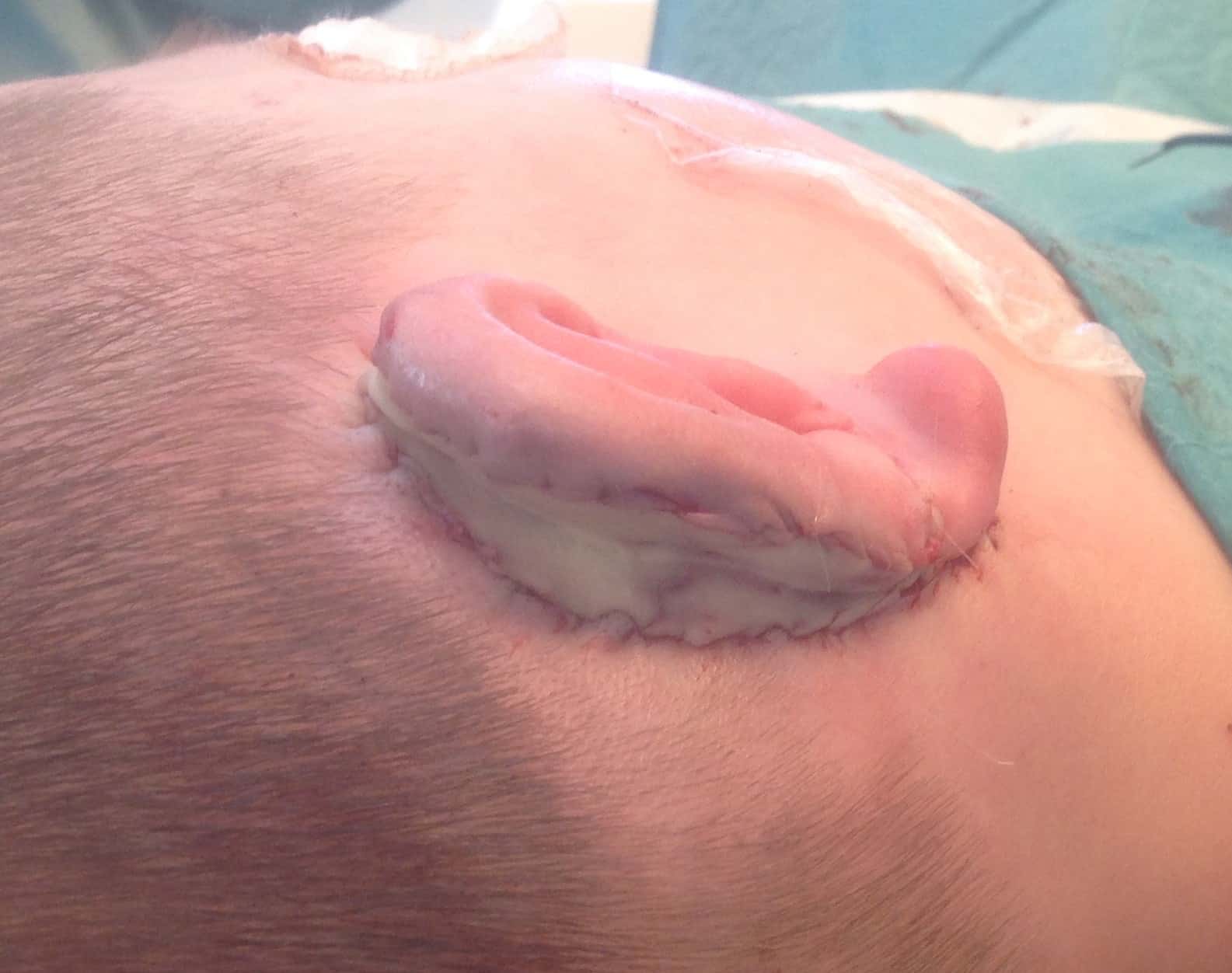
Picture: The rib cartilage ear mould the out ear is reconstructed from (plastic surgeon Jorma Rautio)
Other facial area reconstructions
Facial area tumours like melanoma or basal-cell carcinoma (basaliom) surgeries may require large tissue removals. In these cases, local flaps are used. In wide tissue deficiencies skin graft or microneurovascular free flap might be needed. A temporal fascia flap can be used as a base for skin graft.
Facial nerve paralysis can cause significant asymmetry and dysfunction in the face area which often causes wide social impairment. In reconstructive surgery, the situation can be balanced into a resting position. If the objective is to have mouth movement, a microneurovascular tissue graft is needed. The operation is planned individually depending on the seriousness of the disease.
Full face transplant is the final resort. The first successful full face transplant was performed in Europe many years ago. In Finland, face transplants are carried out in the Helsinki University Hospital.
Facial area reconstructions are demanding and always depend on the quality of the trauma. Plastic surgeon Helena Puonti is specialised in facial corrections. Highly complicated cases might require multiple surgeons from different fields and in these cases, we consult colleagues in those specific fields and arrange the best possible care for each patient.
Facial area tumours
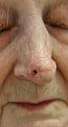
Basal-cell carcinoma is one of the most common facial area tumours. It develops with age usually in areas which have been exposed to sun. It can sometimes appear in younger persons too. Flakiness, redness, and easily cracking skin that does not recover, can be the first signs of basal-cell carcinoma. They should be operated on promptly.
Treatment methods have developed in the past years and a dermatologist can treat superficial redness and cracks with creams or skin peeling. A biopsy of all abnormalities needs to be taken before beginning the cure.
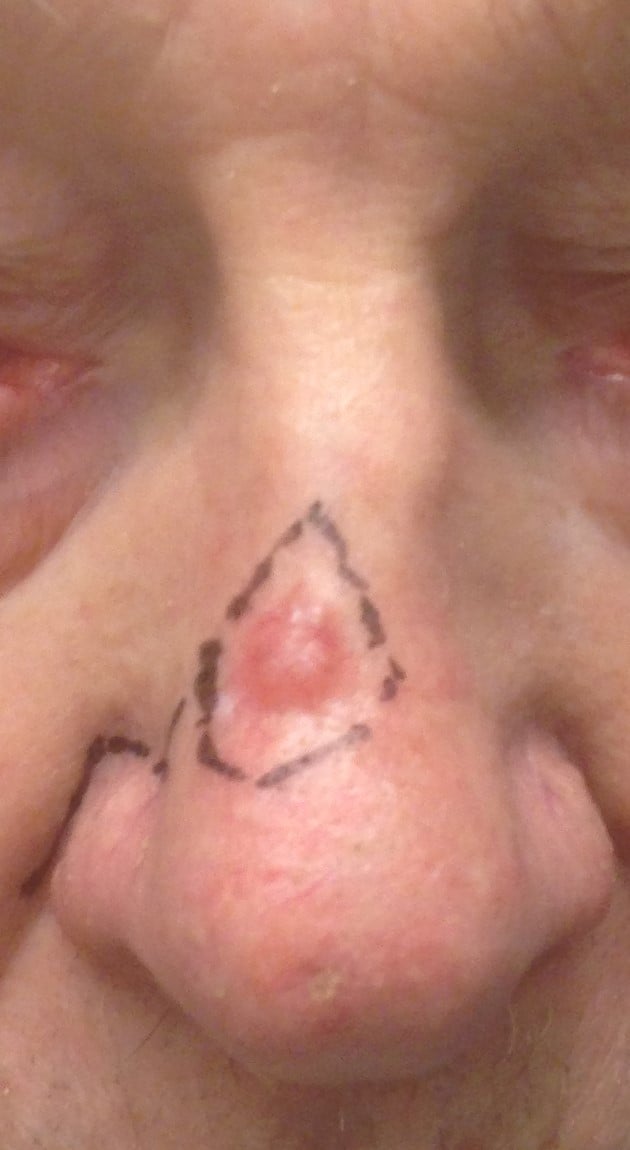
If the skin change has developed into a craterlike, thickened ulcer, surgical removal is the best course of action. In case of basal-cell carcinoma a radical removal with a clear healthy tissue margin is sufficient. The location of the basal-cell carcinoma defines how demanding the operation will be. It can appear in the eye lid and reach the mucus membrane putting the eye at risk. Sometimes the whole eyelid needs to be removed and reconstructed. This can be done with surrounding tissue and if needed, some cartilage can be taken from the ear to support for example the lower lid.
Facial area cancer surgery is highly demanding. Usually, some scarring will remain visible, but we aim to restore the shape when possible.
Other facial area cancers are epidermoid carcinomas present in the surface of the skin, sweat gland carcinoma, melanoma etc. but luckily, these are fairly rare. It is important that of any suspicious abnormality, a biopsy is taken and individual treatment is planned. When suspecting melanoma, the suspicious mole should be wholly removed while taking the biopsy.