Diagnostics
Diagnostics is an important part of breast cancer care and all abnormal symptoms in breasts are worth examining. Any unclear symptoms in a breast should be examined. Methods used to detect breast cancer include clinical examination i.e. palpation of the breast by the doctor, mammography and ultrasound imaging, and a needle biopsy. In the diagnostic phase, analysing the size, location, and tissue sample of the change determine the choice of surgical treatment. Examination of the opposite, “healthy” breast is also carried out.
How is Breast Cancer Diagnosed?
- Clinical breast examination (CBE) is done by a health specialist who palpates the breasts
- Radiologist performs the necessary imaging examinations including breast mammogram and ultrasound
- Unclear changes are subjected to needle biopsy
- Cancer specialist determines the need for a breast MRI
Quick examinations and treatment
If you have already been diagnosed with breast cancer or if, on the basis of your symptoms, you suspect it, you can access Helena Puonti’s consultation quickly and without a referral.
Why choose Clinic Helena:
- World-class expertise in breast cancer treatment, using the most modern methods.
- Helena Puonti’s unique Sensing Breast-method – available only at Clinic Helena.
- We devote ourselves to helping You recover completeley from your illness.
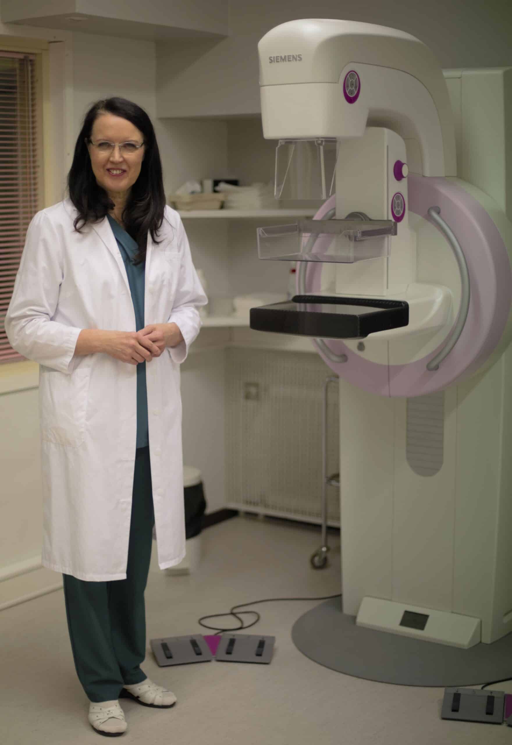
Mammography
Mammography is usually the first method of detecting breast cancer. Mammography can be used to detect changes in the breast, such as the early onset and tissue changes in the early stages of breast cancer.
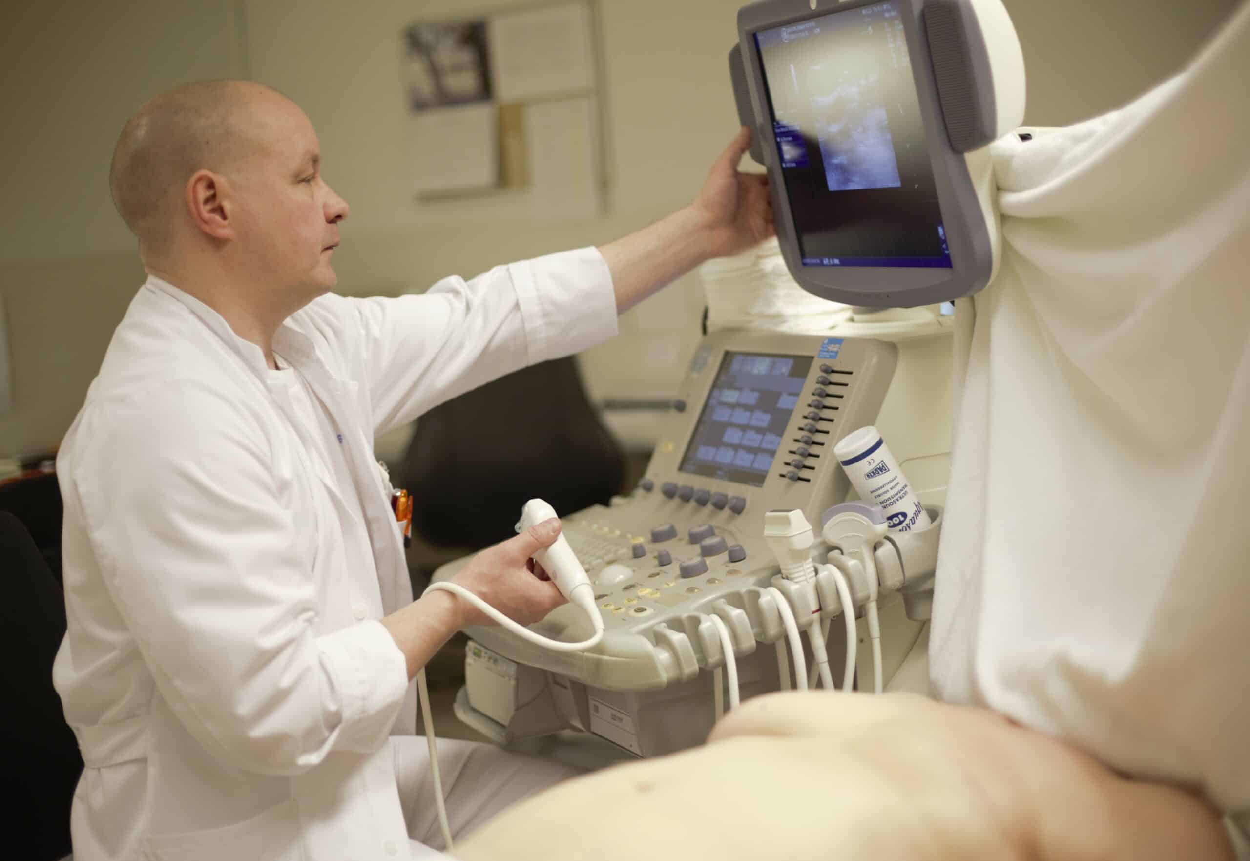
Breast Ultrasound
Breast ultrasound is a method that complements mammography. Ultrasound is a quick and easy procedure and does not cause radiation. In the study, a radiologist will scan the breasts and armpits with ultrasound. During the ultrasound examination, it is also possible to take samples of suspicious changes.
Needle Biopsy
A needle biopsy should be taken from every suspicious change in breast tissue. Histological analysis of the needle biopsy affects the surgeon’s operation plan. A tissue sample of a suspect change is taken by needle biopsy. The tissue sample is usually taken by a radiologist.
Ultrasound or mammography guidance is used for sampling. If necessary, the sample is taken stereotactically under mammography or MRI guidance by needle biopsy (Tru-cut) or vacuum aspiration (vacuum aspiration biopsy).
Breast MRI
If the size or location of the breast change is difficult to determine based on mammography or ultrasound, a breast MRI examination is performed. MRI is also performed if the tumour size and location are unclear, for example in a lobular type of breast cancer, which usually is poorly seen in basic mammography. A radiologist examines the magnetic images of the breasts and gives an opinion on them.

What Happens After a Breast Cancer Diagnosis?
Based on the above-mentioned studies and their results, our surgeon Helena Puonti, who specializes in breast cancer surgery, plans a cancer surgery that is suitable for your disease and aimed at healing you properly so that you can concentrate on returning to your good quality of life.